
neonatal ventilator settings pdf
Neonatal ventilator settings involve adjusting modes and parameters like PEEP, tidal volume, and inspiratory time to support newborns with respiratory distress, ensuring proper gas exchange while minimizing lung injury.
Overview of Neonatal Ventilation
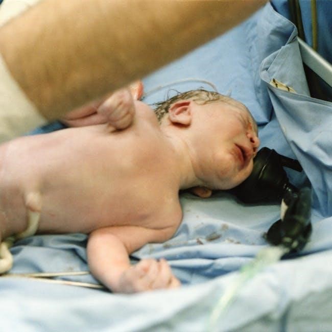
Neonatal ventilation is a critical intervention for newborns with respiratory failure, providing life-saving support by assisting or taking over breathing. It involves delivering air or oxygen through invasive or non-invasive methods, tailored to the infant’s needs. Proper ventilation helps maintain adequate oxygenation, carbon dioxide removal, and lung volume. The goal is to support the neonate’s respiratory system while minimizing complications like lung injury. Various modes, including conventional, advanced, and synchronized ventilation, are used to adapt to the baby’s condition. Effective ventilation requires careful setting adjustments and continuous monitoring to ensure optimal outcomes and prevent long-term pulmonary damage.
Importance of Proper Ventilator Settings
Proper neonatal ventilator settings are critical to ensure effective gas exchange while minimizing lung injury. Incorrect settings can lead to ventilator-induced lung injury (VILI), atelectotrauma, or chronic lung disease. Neonatal lungs are fragile and lack surfactant, making them highly susceptible to damage. Accurate tidal volumes, PEEP levels, and inspiratory times help maintain functional residual capacity, optimizing oxygenation and carbon dioxide removal. Proper settings also reduce the risk of complications, promoting healthier lung development and improving long-term outcomes. Clinicians must carefully balance ventilation support with lung protection, tailoring settings to the infant’s specific condition and response to therapy.
Types of Neonatal Ventilators
Neonatal ventilators are categorized into conventional and advanced types, each designed for specific patient needs. Conventional ventilators provide essential support through modes like assist/control and synchronized intermittent mandatory ventilation (SIMV). Advanced ventilators incorporate specialized modes, such as high-frequency oscillatory ventilation (HFOV) and pressure-support ventilation, offering precise control for critically ill newborns. Some devices also feature non-invasive ventilation options, like nasal continuous positive airway pressure (CPAP), which reduces the need for intubation. Transport ventilators are designed for mobility, ensuring continuous care during patient transfer. Each type is tailored to provide optimal respiratory support while minimizing lung injury, making them essential tools in neonatal intensive care.
Understanding Neonatal Ventilator Modes
Neonatal ventilator modes include conventional, advanced, and synchronized options, each tailored to support respiratory needs. They ensure effective gas exchange while minimizing lung injury, adapting to patient conditions.
Conventional Ventilation Modes
Conventional ventilation modes, such as IPPV (Intermittent Positive Pressure Ventilation) and SIMV (Synchronized Intermittent Mandatory Ventilation), are widely used in neonatal care. These modes deliver breaths at set rates and pressures, providing full or partial respiratory support. IPPV is typically used for newborns who require full ventilation assistance, while SIMV allows the baby to breathe spontaneously between ventilator-delivered breaths, promoting lung function. Initial settings often include PEEP (2-5 cmH2O), PIP (15-25 cmH2O), and a respiratory rate of 30-60 breaths per minute. Proper adjustment of these settings is crucial to avoid lung injury and ensure adequate gas exchange, adapting to the neonate’s evolving respiratory needs for optimal outcomes.
Advanced Ventilation Modes
Advanced ventilation modes, such as HFOV (High-Frequency Oscillatory Ventilation) and Volume-Targeted Ventilation, offer precise control for neonates with severe respiratory distress. HFOV delivers rapid, small tidal volumes to maintain lung recruitment while minimizing injury. It is often used in cases of persistent pulmonary hypertension or severe RDS. Volume-Targeted Ventilation adjusts pressure to ensure consistent tidal volumes, reducing the risk of overdistension. These modes require careful monitoring of lung mechanics and gas exchange. Advanced modes are particularly beneficial for neonates with complex respiratory needs, providing tailored support to optimize outcomes and reduce long-term pulmonary complications. They are typically used in conjunction with other therapies, such as surfactant administration, for enhanced efficacy.
Synchronized Ventilation Modes
Synchronized ventilation modes, such as SIMV (Synchronized Intermittent Mandatory Ventilation) and SIPPV (Synchronized Intermittent Positive Pressure Ventilation), are designed to align with the neonate’s spontaneous breathing efforts. These modes deliver ventilator breaths in synchrony with the baby’s inspirations, promoting a more physiologic breathing pattern. This synchronization reduces asynchrony, enhances comfort, and improves gas exchange. SIMV provides a set rate of mandatory breaths while allowing spontaneous breaths between, while SIPPV synchronizes all ventilator breaths with the patient’s efforts. These modes are particularly beneficial for neonates with some respiratory effort, as they support weaning and reduce the risk of lung injury. Proper synchronization is key to optimizing ventilatory support and patient outcomes.

Initial Ventilator Settings for Neonates
Initial settings are tailored to the neonate’s condition, starting with tidal volumes of 4-6 mL/kg, PIP of 15-25 cmH2O, and PEEP of 4-6 cmH2O to ensure adequate gas exchange and lung recruitment.
Setting Tidal Volume
Tidal volume (VT) is a critical parameter in neonatal ventilation, typically set between 4-6 mL/kg to ensure adequate ventilation while minimizing lung injury. Proper VT helps maintain functional residual capacity, optimizing gas exchange and reducing atelectotrauma. Clinical guidelines emphasize using volume-targeted modes to achieve consistent VT, which is adjusted based on patient response and blood gas analysis. Monitoring lung compliance and resistance helps fine-tune VT for individualized care, ensuring the neonate receives appropriate support without causing ventilator-induced lung injury.
Adjusting Peak Inspiratory Pressure (PIP)
Peak Inspiratory Pressure (PIP) is adjusted to ensure effective ventilation while protecting neonatal lungs from injury. Initial PIP is set based on the neonate’s lung compliance and resistance, typically starting at lower levels to avoid barotrauma. Gradual increments are made to achieve target tidal volumes, ensuring adequate gas exchange. PIP adjustments are guided by clinical assessment, blood gas analysis, and lung mechanics. Using volume-targeted modes helps maintain consistent tidal volumes, while PIP is fine-tuned to prevent excessive pressures. Continuous monitoring is essential to balance oxygenation and ventilation, optimizing outcomes for fragile neonatal patients.
Setting Positive End-Expiratory Pressure (PEEP)
Positive End-Expiratory Pressure (PEEP) is critical in neonatal ventilation to maintain lung volume and prevent alveolar collapse; Initial PEEP levels are typically set between 4-6 cmH2O, depending on the neonate’s gestational age and lung condition. PEEP is adjusted to optimize oxygenation and reduce atelectotrauma, ensuring functional residual capacity is maintained. Blood gas analysis and clinical signs, such as chest rise and breath sounds, guide PEEP adjustments. Higher PEEP may be required for surfactant-deficient lungs, while lower levels are used for more compliant lungs. Fine-tuning PEEP balances oxygenation and ventilation, minimizing lung injury and promoting stable respiratory function in neonates.
Determining Inspiratory Time
Inspiratory time in neonatal ventilation is set to synchronize with the infant’s natural breathing pattern. Typically, it ranges from 0.3 to 0.5 seconds, ensuring adequate gas exchange without causing airway pressure spikes. Adjustments are made based on the infant’s respiratory rate and disease severity. Shorter inspiratory times are used in conditions like respiratory distress syndrome, while longer times may be needed for severe cases to prevent gas trapping. Clinical assessment and continuous monitoring guide inspiratory time optimization, ensuring effective ventilation and minimizing complications like volutrauma or atelectasis, thus promoting lung protection and stable respiratory support for neonates.

Key Parameters in Neonatal Ventilation
Central parameters include respiratory rate, tidal volume, PEEP, and FiO₂, ensuring optimal gas exchange and lung protection. These settings are tailored to the neonate’s specific needs.
Respiratory Rate and Breath Timing
Respiratory rate and breath timing are critical in neonatal ventilation. The rate is typically set between 30-60 breaths per minute to match the neonate’s spontaneous breathing. Breath timing, including inspiratory and expiratory durations, ensures synchronized ventilation. Proper timing prevents breath stacking and asynchrony, reducing the risk of lung injury. Adjustments are made based on the infant’s response, clinical assessment, and blood gas results. Optimal timing supports effective gas exchange while minimizing barotrauma and volutrauma. Continuous monitoring ensures that these parameters remain aligned with the neonate’s evolving respiratory needs, promoting lung protection and overall clinical stability.
Fraction of Inspired Oxygen (FiO2)
Fraction of Inspired Oxygen (FiO2) is a crucial parameter in neonatal ventilation, determining the percentage of oxygen delivered to the newborn. It is typically set between 21% and 100%, adjusted to maintain target oxygen saturation levels. Proper FiO2 titration prevents hypoxia and hyperoxia, both of which can cause significant complications. Clinicians closely monitor oxygen saturation via pulse oximetry and adjust FiO2 accordingly. Blood gas analysis also guides FiO2 adjustments, ensuring optimal oxygenation without increasing the risk of oxidative stress or retinopathy of prematurity. Maintaining the correct FiO2 balance is essential for supporting the neonate’s respiratory health and overall development.
Flow Rates and Their Implications
Flow rates in neonatal ventilation refer to the speed of gas delivery during inspiration, typically measured in liters per minute (L/min). Proper flow rate settings ensure synchronized breathing with the ventilator, preventing asynchrony and discomfort. Higher flow rates may be needed for neonates with increased respiratory resistance or distress, while lower rates are used for smaller or less active patients. Incorrect flow rates can lead to inadequate gas exchange or increased work of breathing, potentially causing fatigue or lung injury; Monitoring flow rates alongside clinical responses is essential for optimizing ventilation support and ensuring the neonate’s respiratory needs are met effectively.

Monitoring Neonatal Ventilator Settings
Clinical assessment, blood gas analysis, and continuous sensor monitoring are crucial for evaluating ventilator effectiveness, ensuring proper oxygenation, and guiding adjustments to maintain optimal respiratory support for neonates.
Clinical Assessment of Ventilation
Clinical assessment of ventilation in neonates involves evaluating respiratory mechanics, oxygenation, and overall patient response. Observing chest movement, breath sounds, and respiratory rate helps ensure effective ventilation. Pulse oximetry monitors oxygen saturation, while capillary blood gas analysis provides insights into carbon dioxide levels and acid-base balance. Clinical signs such as retractions, grunting, or nasal flaring indicate respiratory distress. Regular assessment of lung compliance and resistance guides adjustments in ventilator settings. Additionally, chest X-rays are used to confirm endotracheal tube placement and detect complications like atelectasis or pneumothorax. Continuous clinical monitoring ensures ventilatory support is optimized, promoting lung protection and improving outcomes for neonates.
Using Blood Gas Analysis
Blood gas analysis is a critical tool in neonatal ventilation, providing insights into oxygenation and ventilation status. It measures pH, PaCO2, and PaO2 levels, helping to assess the effectiveness of ventilator settings. By identifying acid-base imbalances, clinicians can adjust parameters like FiO2, PEEP, and respiratory rate to optimize gas exchange. Blood gas results guide titration of oxygen therapy and mechanical ventilation, ensuring adequate oxygen delivery while minimizing the risk of hyperoxia or hypercapnia. Regular analysis helps in fine-tuning ventilator settings, promoting lung healing, and preventing complications. This data-driven approach ensures personalized and effective respiratory support for neonates, improving clinical outcomes and reducing morbidity.
Continuous Monitoring with Sensors
Continuous monitoring with sensors is essential in neonatal ventilation to ensure real-time assessment of respiratory parameters. Sensors integrated into ventilators track tidal volumes, inspiratory pressures, and flow rates, providing immediate feedback on the effectiveness of ventilation. This allows clinicians to detect subtle changes in the neonate’s condition, such as air leaks or disconnections, and make prompt adjustments. Advanced sensors also monitor carbon dioxide levels (capnography) and oxygen saturation (SpO2), offering a comprehensive view of the neonate’s respiratory and cardiovascular status. Continuous data collection enhances patient-ventilator synchrony, reduces complications, and supports timely interventions, ensuring safer and more effective respiratory support for critically ill newborns.

Complications of Neonatal Ventilation
Complications of neonatal ventilation include ventilator-induced lung injury (VILI), atelectotrauma, and chronic lung disease (CLD), emphasizing the need for precise settings to protect fragile neonatal lungs.
Ventilator-Induced Lung Injury (VILI)
Ventilator-induced lung injury (VILI) is a significant complication in neonatal ventilation, caused by mechanical stress from excessive pressure or volume, leading to inflammation and lung tissue damage. Overdistension of alveoli and repetitive collapse/reopening of airways contribute to this injury. VILI can result in prolonged ventilation, chronic lung disease, and long-term respiratory morbidity. Pathophysiology involves release of inflammatory mediators and disruption of the alveolar-capillary barrier. Clinicians must balance adequate ventilation with lung protection to minimize VILI risk, emphasizing the importance of careful ventilator settings and monitoring. Strategies like low tidal volumes and optimal PEEP are critical to mitigate this injury in vulnerable neonates.
Atelectotrauma and Lung Compliance
Atelectotrauma refers to lung damage caused by the repetitive collapse and reopening of alveoli during mechanical ventilation. Neonatal lungs, being highly compliant and surfactant-deficient, are particularly susceptible. Low lung volumes and insufficient positive end-expiratory pressure (PEEP) exacerbate alveolar collapse, leading to shear stress and inflammation. Optimizing PEEP settings is crucial to maintain functional residual capacity (FRC) and prevent atelectotrauma. Improved lung compliance enhances gas exchange efficiency and reduces the risk of chronic lung disease. Clinicians must carefully titrate ventilator pressures to balance lung recruitment and overdistension, ensuring that the delicate neonatal lungs are protected throughout the ventilation process. This balance is vital for minimizing long-term respiratory complications.
Chronic Lung Disease (CLD) Risks
Chronic Lung Disease (CLD), also known as bronchopulmonary dysplasia (BPD), is a significant complication of neonatal mechanical ventilation. Prolonged ventilation with inappropriate settings can damage developing lungs, leading to long-term respiratory and neurodevelopmental issues. Overdistension and atelectotrauma from excessive pressures and inadequate PEEP contribute to lung injury. Premature infants, especially those with low birth weight, are at higher risk. Optimizing ventilator settings to maintain lung recruitment while avoiding excessive tidal volumes is critical. Early identification of CLD risks and tailored ventilatory strategies can mitigate complications, improving long-term outcomes for vulnerable neonates. Clinicians must balance ventilation support with lung protection to minimize CLD risks effectively.

Adjusting Ventilator Settings
Adjusting ventilator settings involves titrating FiO2, gradually weaning support, and responding to clinical changes to optimize oxygenation and ventilation while minimizing lung injury in neonates.
Titration of FiO2
Titrating FiO2 involves adjusting the fraction of inspired oxygen to maintain target oxygen saturation levels, typically between 90-95%, to prevent hypoxia and hyperoxia. This process requires continuous monitoring of arterial oxygen levels and clinical assessment of the neonate’s condition. The goal is to use the lowest FiO2 necessary to achieve optimal oxygenation while minimizing the risk of oxidative stress and retinopathy of prematurity; Clinicians must balance oxygenation needs with potential risks, ensuring that changes in FiO2 are gradual and guided by blood gas analysis and pulse oximetry readings. Regular adjustments are crucial to support the neonate’s evolving respiratory needs.
Weaning strategies aim to gradually reduce mechanical ventilation support as the neonate’s respiratory function improves. This process involves lowering the respiratory rate, decreasing inspiratory pressure, and increasing the use of synchronized ventilation modes. The goal is to transition the neonate to self-breathing while ensuring stability. Clinicians monitor parameters like tidal volume, respiratory effort, and arterial blood gases to guide the weaning process. Successful weaning requires careful assessment of the neonate’s readiness and gradual adjustments to avoid respiratory fatigue or distress. Protocols often include stepwise reductions in ventilator settings, ensuring a smooth transition to independent breathing. Regular monitoring and clinical judgment are key to effective weaning. Response to clinical deterioration in neonates requires prompt adjustment of ventilator settings to stabilize respiratory function. Clinicians must evaluate oxygenation and carbon dioxide levels, assessing for signs of distress or malfunction. Increasing FiO2 or PIP may be necessary to improve oxygenation, while ensuring tidal volumes remain within safe limits to prevent lung injury. Continuous monitoring of vital signs and blood gases guides interventions. Synchronized ventilation modes can enhance breath timing, reducing asynchrony. If deterioration persists, advanced therapies like surfactant administration or changing ventilation modes may be considered. Timely recognition and intervention are critical to prevent further complications and ensure optimal neonatal outcomes. Regular reassessment is essential. Clinical guidelines standardize neonatal ventilator care, ensuring evidence-based practices are followed. Protocols outline step-by-step procedures for setting and adjusting ventilators, while respiratory teams provide specialized support, optimizing outcomes for critically ill newborns. Evidence-based practices in neonatal ventilation are grounded in clinical trials and expert consensus, ensuring safe and effective respiratory support. Guidelines recommend lung-protective strategies, such as using appropriate PEEP levels and tidal volumes, to minimize lung injury. Surfactant therapy is a cornerstone for preterm infants with respiratory distress syndrome, supported by robust evidence. Ventilator settings should be tailored to the infant’s weight, gestational age, and underlying condition. Regular monitoring of blood gas results and clinical status guides adjustments. These practices aim to optimize oxygenation, reduce complications, and improve long-term outcomes, emphasizing a balanced approach to ventilation that prioritizes both efficacy and safety for vulnerable newborns. Protocols for ventilator management in neonates are standardized procedures designed to ensure consistent and effective respiratory support. These protocols typically include guidelines for initiating ventilation, selecting appropriate modes, and adjusting settings based on patient response. Initial assessments involve determining the infant’s gestational age, weight, and underlying condition to set baseline parameters. Continuous monitoring of vital signs, blood gas results, and clinical status is essential to guide adjustments. Protocols also emphasize the importance of documenting changes and ensuring interdisciplinary collaboration. Regular reviews and updates to protocols are necessary to align with evolving evidence and improve patient outcomes, ensuring safe and effective ventilator use. Neonatal respiratory teams play a critical role in managing ventilator settings, ensuring optimal respiratory support for critically ill newborns. These teams, comprising neonatologists, respiratory therapists, and nurses, collaborate to assess each infant’s unique needs. They interpret clinical data, including blood gas results and imaging, to adjust ventilator parameters. Their expertise ensures that settings like tidal volume, PEEP, and FiO2 are tailored to minimize lung injury while maintaining adequate oxygenation. Regular rounds and continuous monitoring allow the team to respond promptly to changes in the infant’s condition. Their coordinated efforts aim to improve outcomes, reduce complications, and facilitate a smooth transition to less invasive support as the infant recovers. Special considerations include surfactant therapy, non-invasive ventilation techniques, and safe transport strategies for neonates on ventilators, ensuring tailored support while minimizing complications and optimizing outcomes. Surfactant therapy is a critical intervention for preterm neonates with respiratory distress syndrome (RDS). It involves administering exogenous surfactant to reduce surface tension in the lungs, improving gas exchange and lung compliance. Surfactant is typically given via an endotracheal tube and is most effective when administered early in the course of respiratory failure. It decreases the work of breathing and reduces the need for high ventilator pressures, minimizing the risk of lung injury. Clinical guidelines recommend surfactant therapy for neonates with RDS, as it has been shown to improve outcomes and reduce mortality. Proper timing and dosage are essential for optimal benefits. Non-invasive ventilation (NIV) techniques are increasingly used in neonatal care to support breathing without requiring intubation. Continuous positive airway pressure (CPAP) is the most common method, providing a constant pressure to keep airways open. Biphasic positive airway pressure (BiPAP) alternates between two pressure levels, aiding spontaneous breathing. High-flow nasal cannula (HFNC) delivers heated, humidified gas at high flow rates, improving oxygenation and reducing work of breathing. These techniques are particularly beneficial for preterm infants with respiratory distress syndrome (RDS) or mild-to-moderate respiratory failure. NIV minimizes the risk of ventilator-induced lung injury and promotes earlier weaning from mechanical support, improving long-term pulmonary outcomes. Transporting neonates on ventilators requires careful planning and specialized equipment to ensure continued respiratory support during movement. Portable ventilators designed for transport are used, often equipped with modes like CPAP and synchronized ventilation to maintain stability. Key parameters such as FiO2, PEEP, and respiratory rate must be closely monitored. The transport team must be trained to manage potential complications like disconnections or power failures. Regular checks of ventilator settings and battery life are essential before and during transport. This ensures the neonate receives uninterrupted care, minimizing risks and maintaining clinical stability throughout the transfer process. Troubleshooting neonatal ventilator issues involves identifying equipment malfunction, managing air leaks, and addressing patient-ventilator asynchrony. Promptly resolving these ensures continuous, effective respiratory support for neonates. Identifying equipment malfunction in neonatal ventilators is critical for ensuring patient safety and effective ventilation. Clinicians must monitor for alarms, unusual sounds, or inconsistent pressure readings. Regular checks of tubing, connections, and sensors are essential to detect disconnections or blockages. Comparing ventilator settings with clinical observations, such as chest rise and breath sounds, helps verify functionality. If malfunction is suspected, immediate troubleshooting, including resetting or replacing the device, is necessary. Adhering to clinical guidelines and manufacturer protocols ensures timely resolution and minimizes risks to the neonate. Proper maintenance and staff training are key to preventing and managing equipment issues effectively. Managing air leaks and disconnections is crucial in neonatal ventilation to prevent ineffective gas exchange and lung injury. Air leaks often occur due to improper endotracheal tube positioning or uncuffed tubes, which can lead to loss of delivered tidal volume. Clinicians must ensure secure connections and regularly inspect tubing for damage or disconnections. Using cuffed endotracheal tubes and securing devices can minimize leaks. If a leak is detected, adjusting the ventilator settings, such as increasing flow or inspiratory time, may help compensate. Continuous monitoring of ventilator alarms and clinical signs, like decreased breath sounds or chest rise, is essential for early detection and intervention. Patient-VENT asynchrony occurs when a neonate’s spontaneous breathing does not align with the ventilator’s delivered breaths, reducing efficiency and increasing lung stress. To address this, clinicians should monitor breath patterns using flow and pressure waveforms. Adjusting ventilator settings, such as inspiratory time or trigger sensitivity, can improve synchronization. Synchronized ventilation modes, like SIMV or A/C, are beneficial as they align mechanical breaths with the patient’s efforts. If asynchrony persists, consider changing the ventilation mode or adding non-invasive support. Regular clinical assessment and adjustments are critical to ensure effective ventilation and minimize complications. Best practices for neonatal ventilator settings emphasize evidence-based approaches to balance oxygenation, ventilation, and lung protection. Use of non-invasive ventilation when possible, synchronization with the infant’s breaths, and careful titration of FiO₂ are key. Monitoring with blood gas analysis and clinical assessments ensures optimal settings. Regular adjustments based on patient response and lung mechanics are essential. Surfactant therapy and prone positioning can enhance outcomes. Weaning strategies should be gradual, with close monitoring for signs of respiratory distress. Collaborative efforts between neonatologists, respiratory therapists, and nurses are critical for achieving the best results and minimizing long-term complications. Recent advancements in neonatal ventilation technology include the development of microprocessor-controlled ventilators, enabling precise adjustments and real-time monitoring. Servo-controlled ventilators now offer synchronized breaths with the patient, reducing asynchrony. Volume-targeted modes ensure consistent tidal volumes, while closed-loop systems automatically adjust settings based on feedback. Neurally adjusted ventilatory assist (NAVA) uses electrical activity from the diaphragm to synchronize support. Portable ventilators with advanced modes enhance transport capabilities. These innovations improve lung protection, reduce complications, and enhance clinical outcomes. Integration with non-invasive techniques like nasal CPAP and surfactant therapy further minimizes invasive interventions, promoting gentler respiratory support for vulnerable newborns.Weaning Strategies
Response to Clinical Deterioration
Clinical Guidelines and Protocols
Evidence-Based Practices
Protocols for Ventilator Management
Role of Neonatal Respiratory Teams

Special Considerations in Neonatal Ventilation
Surfactant Therapy
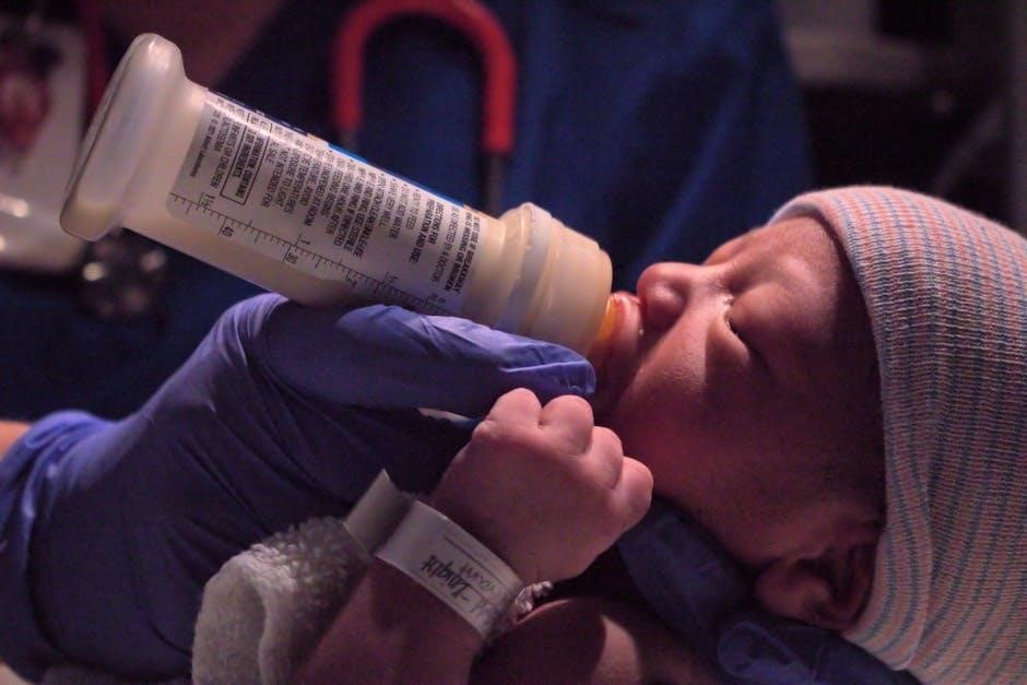
Non-Invasive Ventilation Techniques
Transporting Neonates on Ventilators

Troubleshooting Neonatal Ventilator Issues
Identifying Equipment Malfunction
Managing Air Leaks and Disconnections
Addressing Patient-VENT Asynchrony
Advancements in Neonatal Ventilation Technology